Parenchymal Infections: Prions
![]()
Parenchymal Infections: Prions
![]()
Scrapie is a chronic disease of sheep which is transmitted by a filterable particle that is resistant to heat and formalin fixation. The human disease kuru, which is also caused by a filterable particle, is found in the Fore speaking people of New Guinea. The disease is perpetuated through the practice of cannibalism. These people eat the bodies of their own dead. Because only the women and children eat the brains, kuru is usually not found in the adult male population. Having discovered the relationship between cannibalism and kuru, the practice of cannibalism has been stopped and the incidence of the disease has abated. Kuru has a slow onset and is manifested by cerebellar signs and shaking ataxia. Death occurs with 18 to 24 months.
Until recently, the infective agents of kuru and Creutzfeldt-Jakob disease in humans, and scrapie in sheep, were presumed to be slow, latent viruses. The latest hypothesis is that these diseases are caused by prions. Prions are composed of protein and are much smaller than viruses.
Kuru
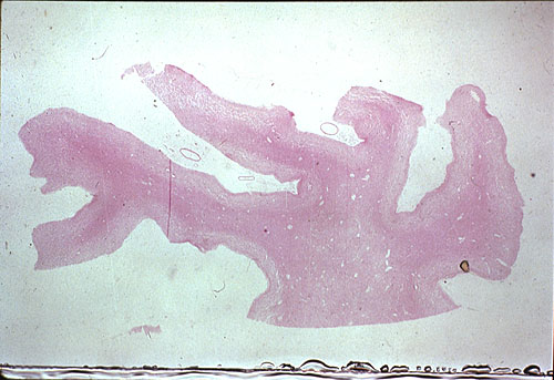
![]() The
gross findings in kuru are minimal, and consist of congestion of blood
vessels, which can be seen on this slide, and in long standing cases, cortical
atrophy (not prominent in this case).
The
gross findings in kuru are minimal, and consist of congestion of blood
vessels, which can be seen on this slide, and in long standing cases, cortical
atrophy (not prominent in this case).
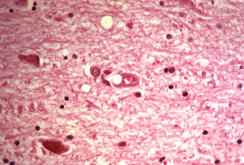
![]() Microscopically,
one finds vacuolization in neuronal cytoplasm and dendrites, which gives
the neuropil a spongy appearance.
Microscopically,
one finds vacuolization in neuronal cytoplasm and dendrites, which gives
the neuropil a spongy appearance.
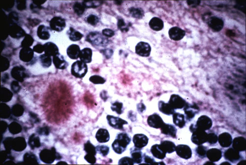
![]() Another
characteristic finding of kuru is shown on this slide: brush-like
plaques, which are also called spikeballs and found in the cerebellum.
Another
characteristic finding of kuru is shown on this slide: brush-like
plaques, which are also called spikeballs and found in the cerebellum.
Creutzfeldt-Jakob disease
Creutzfeldt-Jakob disease is an uncommon, sporadic, degenerative disease in humans, which usually occurs during the middle decades of life. The course is subacute, ranging from 3 to 6 months in duration, and is manifested by dementia accompanied by motor signs and blindness. The infective agent can be transmitted experimentally to chimpanzees. It is noteworthy that upon its initial passage the infective agent exists of a particle having a diameter of 120 to 150 µm. With subsequent passages in chimps, the particle becomes much smaller and the incubation period becomes shorter.
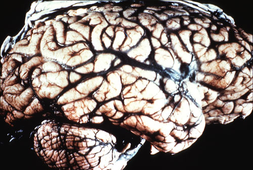
![]() The
gross pathology of Creutzfeldt-Jakob disease is that of cortical atrophy,
manifest here by marked widening of sulci, and gliosis.
The
gross pathology of Creutzfeldt-Jakob disease is that of cortical atrophy,
manifest here by marked widening of sulci, and gliosis.
![]() Microscopically,
spongy changes similar to those seen in kuru are found in the cortex. It
is paramount when making the diagnosis of Creutzfeldt-Jakob disease, that
one is able to demonstrate that at least some of the vacuoles arise within
neuronal cytoplasm. Other changes usually seen include red neurons and
gliosis.
Microscopically,
spongy changes similar to those seen in kuru are found in the cortex. It
is paramount when making the diagnosis of Creutzfeldt-Jakob disease, that
one is able to demonstrate that at least some of the vacuoles arise within
neuronal cytoplasm. Other changes usually seen include red neurons and
gliosis.